genevalley's北美文摘
最前沿的生命科學,包括發現,思想和文章。 最貼近的北美生活,包括科研,生活和綠卡。


| |||||||||||||||||||||||||||||||||


|
|
| Previous Article | Next Article |
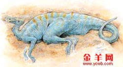
Soft-Tissue Vessels and Cellular Preservation in Tyrannosaurus rex

Soft tissues are preserved within hindlimb elements of Tyrannosaurus rex (Museum of the Rockies specimen 1125). Removal of the mineral phase reveals transparent, flexible, hollow blood vessels containing small round microstructures that can be expressed from the vessels into solution. Some regions of the demineralized bone matrix are highly fibrous, and the matrix possesses elasticity and resilience. Three populations of microstructures have cell-like morphology. Thus, some dinosaurian soft tissues may retain some of their original flexibility, elasticity, and resilience.
1 Department of Marine, Earth, Atmospheric Sciences, North Carolina State University, Raleigh, NC 27695, USA.
2 North Carolina State Museum of Natural Sciences, Raleigh, NC 27601, USA.
3 Museum of the Rockies, Montana State University, Bozeman, MT 59717, USA.
4 Carnegie Institution of Washington, Geophysical Laboratory, 5251 Broad Branch Road N.W., Washington, DC 20018, USA.
 Present address: Department of Geosciences, Christian-Albrechts University Kiel, Olshausenstrasse 40, 24098 Kiel, Germany.
 ![]()
* To whom correspondence should be addressed. E-mail: schweitzer@ncsu.edu.
A newly discovered specimen of Tyrannosaurus rex [Museum of the Rockies (MOR) specimen 1125] was found at the base of the Hell Creek Formation, 8 m above the Fox Hills Sandstone, as an association of disarticulated elements. The specimen was incorporated within a soft, well-sorted sandstone that was interpreted as estuarine in origin. Although some bones are slightly deformed or crushed, preservation is excellent. MOR 1125 represents a relatively small individual of T. rex, with a femoral length of 107 cm, as compared to the Field Museum (Chicago) specimen (FMNH PR2081) that has a femoral length of approximately 131 cm. On the basis of calculated lines of arrested growth (LAG), we estimated that this animal was 18 ± 2 years old at death (1).
No preservatives were applied to interior fragments of the femur of MOR 1125 during preparation, and these fragments were reserved for chemical analyses. In addition to the dense compact bone typical of theropods, this specimen contained regions of unusual bone tissue on the endosteal surface (2). Cortical and endosteal bone tissues were demineralized (3), and after 7 days, several fragments of the lining tissue exhibited unusual characteristics not normally observed in fossil bone. Removal of the mineral phase left a flexible vascular tissue that demonstrated great elasticity and resilience upon manipulation. In some cases, repeated stretching was possible (Fig. 1A, arrow), and small pieces of this demineralized bone tissue could undergo repeated dehydration-rehydration cycles (Fig. 1B) and still retain this elastic character. Demineralization also revealed that some regions of the bone were highly fibrous (Fig. 1C, arrows).
 Fig. 1. Demineralized fragments of endosteally derived tissues lining the marrow cavity of the T. rex femur. (A) The demineralized fragment is flexible and resilient and, when stretched (arrow), returns to its original shape. (B) Demineralized bone in (A) after air drying. The overall structural and functional characteristics remain after dehydration. (C) Regions of demineralized bone show fibrous character (arrows). Scale bars, 0.5 mm. Fig. 1. Demineralized fragments of endosteally derived tissues lining the marrow cavity of the T. rex femur. (A) The demineralized fragment is flexible and resilient and, when stretched (arrow), returns to its original shape. (B) Demineralized bone in (A) after air drying. The overall structural and functional characteristics remain after dehydration. (C) Regions of demineralized bone show fibrous character (arrows). Scale bars, 0.5 mm. |
Partial demineralization of the cortical bone revealed parallel-oriented vascular canals that were seen to bifurcate in some areas (Fig. 2A, arrows). Occasional fenestrae (marked F) were observed on the surface of the vascular canals, possibly correlating with communicating Volkmann's canals. Complete demineralization of the cortical bone released thin and transparent soft-tissue vessels from some regions of the matrix (Fig. 2, B and C), which floated freely in the demineralizing solution. Vessels similar in diameter and texture were recovered from extant ostrich bone, when demineralization was followed by digestion with collagenase enzyme (3) to remove densely fibrous collagen matrix (Fig. 2D). In both dinosaur (Fig. 2C) and ostrich (Fig. 2D), remnants of the original organic matrix in which the vessels were embedded can still be visualized under transmitted light microscopy. These vessels are flexible, pliable, and translucent (Fig. 2E). The vessels branch in a pattern consistent with extant vessels, and many bifurcation points are visible (Fig. 2E, arrows). Many of the dinosaur vessels contain small round microstructures that vary from deep red to dark brown (Fig. 2, F and G). The vessels and contents are similar in all respects to blood vessels recovered from extant ostrich bone (Fig. 2H). Aldehyde-fixed (3) dinosaur vessels (Fig. 2I) are virtually identical in overall morphology to similarly prepared ostrich vessels (Fig. 2J), and structures consistent with remnants of nuclei from the original endothelial cells are visible on the exterior of both dinosaur and ostrich specimens (Fig. 2, I and J, arrows).
 Fig. 2. Demineralization of cortical bone reveals the presence of soft-tissue structures. (A) Partial demineralization of a fragment of T. rex cortical bone shows an emerging network of vascular canals, some of which are bifurcated (arrows). All are aligned in parallel, consistent with Haversian canals in cortical bone. Small fenestrae (marked F) may indicate invaginations for communicating Volkmann's canals. (B) A second fragment of T. rex cortical bone illustrates transparent vessels (arrows) arising from bone matrix in solution. (C) Complete demineralization reveals transparent flexible vessels in what remains of the cortical bone matrix, represented by a brown amorphous substance (marked M). (D) Ostrich vessel after demineralization of cortical bone and subsequent digestion of fibrous collagenous matrix. Transparent vessels branch and remain associated with small regions of undigested bone matrix, seen here as amorphous, white fibrous material (marked M). Scale bars in (A) to (D), 0.5 mm. (E) Higher magnification of dinosaur vessels shows branching pattern (arrows) and internal contents. Vascular structure is not consistent with fungal hyphae (no septae, and branching pattern is not consistent with fungal morphology) or plant (no cell walls visible, and again branching pattern is not consistent). Round red microstructures within the vessels are clearly visible. (F) T. rex vessel fragment, containing microstructures consistent in size and shape with those seen in the ostrich vessel in (H). (G) Second fragment of dinosaur vessel. Air/fluid interfaces, represented by dark menisci, illustrate the hollow nature of vessels. Microstructure is visible within the vessel. (H) Ostrich vessel digested from demineralized cortical bone. Red blood cells can be seen inside the branching vessel. (I) T. rex vessel fragment showing detail of branching pattern and structures morphologically consistent with endothelial cell nuclei (arrows) in vessel wall. (J) Ostrich blood vessel liberated from demineralized bone after treatment with collagenase shows branching pattern and clearly visible endothelial nuclei. Scale bars in (E) to (J), 50 µm. (F), (I), and (J) were subjected to aldehyde fixation (3). The remaining vessels are unfixed. Fig. 2. Demineralization of cortical bone reveals the presence of soft-tissue structures. (A) Partial demineralization of a fragment of T. rex cortical bone shows an emerging network of vascular canals, some of which are bifurcated (arrows). All are aligned in parallel, consistent with Haversian canals in cortical bone. Small fenestrae (marked F) may indicate invaginations for communicating Volkmann's canals. (B) A second fragment of T. rex cortical bone illustrates transparent vessels (arrows) arising from bone matrix in solution. (C) Complete demineralization reveals transparent flexible vessels in what remains of the cortical bone matrix, represented by a brown amorphous substance (marked M). (D) Ostrich vessel after demineralization of cortical bone and subsequent digestion of fibrous collagenous matrix. Transparent vessels branch and remain associated with small regions of undigested bone matrix, seen here as amorphous, white fibrous material (marked M). Scale bars in (A) to (D), 0.5 mm. (E) Higher magnification of dinosaur vessels shows branching pattern (arrows) and internal contents. Vascular structure is not consistent with fungal hyphae (no septae, and branching pattern is not consistent with fungal morphology) or plant (no cell walls visible, and again branching pattern is not consistent). Round red microstructures within the vessels are clearly visible. (F) T. rex vessel fragment, containing microstructures consistent in size and shape with those seen in the ostrich vessel in (H). (G) Second fragment of dinosaur vessel. Air/fluid interfaces, represented by dark menisci, illustrate the hollow nature of vessels. Microstructure is visible within the vessel. (H) Ostrich vessel digested from demineralized cortical bone. Red blood cells can be seen inside the branching vessel. (I) T. rex vessel fragment showing detail of branching pattern and structures morphologically consistent with endothelial cell nuclei (arrows) in vessel wall. (J) Ostrich blood vessel liberated from demineralized bone after treatment with collagenase shows branching pattern and clearly visible endothelial nuclei. Scale bars in (E) to (J), 50 µm. (F), (I), and (J) were subjected to aldehyde fixation (3). The remaining vessels are unfixed. |
Under scanning electron microscopy (SEM) (Fig. 3), features seen on the external surface of dinosaurian vessels are virtually indistinguishable from those seen in similarly prepared extant ostrich vessels (Fig. 3, B and F), suggesting a common origin. These features include surface striations that may be consistent with endothelial cell junctions, or alternatively may be artifacts of fixation and/or dehydration. In addition, small round to oval features dot the surface of both dinosaur and ostrich vessels, which may be consistent with endothelial cell nuclei (Fig. 3, E and F, arrows).
 Fig. 3. SEM images of aldehyde-fixed vessels. (A) Isolated vessel from T. rex. (B) Vesselisolated from extant ostrich after demineralization and collagenase digestion (3). (C) Vesselfrom T. rex, showing internal contents and hollow character. (D) Exploded T. rex vessel showing small round microstructures partially embedded in internal vessel walls. (E) Highermagnification of a portion of T. rex vessel wall, showing hypothesized endothelial nuclei (EN). (F) Similar structures visible on fixed ostrich vessel. Striations are seen in both (E) and (F) that may represent endothelial cell junctions or alternatively may be artifacts of the fixation/dehydration process. Scale bars in (A) and (B), 40 µm; in (C) and (D), 10 µm; in (E) and (F), 1 µm. Fig. 3. SEM images of aldehyde-fixed vessels. (A) Isolated vessel from T. rex. (B) Vesselisolated from extant ostrich after demineralization and collagenase digestion (3). (C) Vesselfrom T. rex, showing internal contents and hollow character. (D) Exploded T. rex vessel showing small round microstructures partially embedded in internal vessel walls. (E) Highermagnification of a portion of T. rex vessel wall, showing hypothesized endothelial nuclei (EN). (F) Similar structures visible on fixed ostrich vessel. Striations are seen in both (E) and (F) that may represent endothelial cell junctions or alternatively may be artifacts of the fixation/dehydration process. Scale bars in (A) and (B), 40 µm; in (C) and (D), 10 µm; in (E) and (F), 1 µm. |
Finally, in those regions of the bone where fibrillar matrix predominated in the demineralized tissues, elongate microstructures could be visualized among the fibers (Fig. 4A, inset). These microstructures contain multiple projections on the external surface and are virtually identical in size, location, and overall morphology to osteocytes seen among collagen fibers of demineralized ostrich bone (Fig. 4B, inset). These cell-like microstructures could be isolated and, when subjected to aldehyde fixation (3), appeared to possess internal contents (Fig. 4C), including possible nuclei (Fig. 4C, inset). These microstructures are similar in morphology to fixed ostrich osteocytes, both unstained (Fig. 4D) and stained (3) for better visualization (Fig. 4D, inset). SEM verifies the presence of the features seen in transmitted light microscopy, and again, projections extending from the surface of the microstructures are clearly visible (Fig. 4, E and F).
 Fig. 4. Cellular features associated with T. rex and ostrich tissues. (A) Fragment of demineralized cortical bone from T. rex, showing parallel-oriented fibers and cell-like microstructures among the fibers. The inset is a higher magnification of one of the microstructures seen embedded in the fibrous material. (B) Demineralized and stained (3) ostrich cortical bone, showing fibrillar, parallel-oriented collagen matrix with osteocytes embedded among the fibers. The inset shows a higher magnification of one of the osteocytes. Both inset views show elongate bodies with multiple projections arising from the external surface consistent with filipodia. (C) Isolated microstructure from T. rex after fixation. In addition to the multiple filipodial-like projections, internal contents can be seen. The inset shows a second structure with long filipodia and an internal transparent nucleus-like structure. (D) Fixed ostrich osteocyte; inset, ostrich osteocyte fixed and stained for better visualization. Internal contents are discernible, and filipodia can be seen extending in multiple planes from the cell surface. (E and F) SEM images of aldehyde-fixed (3) microstructures isolated from T. rex cortical bone tissues. Scale bars in (A) and (B), 50 µm; in (C) and (D), 20 µm; in (E), 10 µm; in (F), 1 µm. Fig. 4. Cellular features associated with T. rex and ostrich tissues. (A) Fragment of demineralized cortical bone from T. rex, showing parallel-oriented fibers and cell-like microstructures among the fibers. The inset is a higher magnification of one of the microstructures seen embedded in the fibrous material. (B) Demineralized and stained (3) ostrich cortical bone, showing fibrillar, parallel-oriented collagen matrix with osteocytes embedded among the fibers. The inset shows a higher magnification of one of the osteocytes. Both inset views show elongate bodies with multiple projections arising from the external surface consistent with filipodia. (C) Isolated microstructure from T. rex after fixation. In addition to the multiple filipodial-like projections, internal contents can be seen. The inset shows a second structure with long filipodia and an internal transparent nucleus-like structure. (D) Fixed ostrich osteocyte; inset, ostrich osteocyte fixed and stained for better visualization. Internal contents are discernible, and filipodia can be seen extending in multiple planes from the cell surface. (E and F) SEM images of aldehyde-fixed (3) microstructures isolated from T. rex cortical bone tissues. Scale bars in (A) and (B), 50 µm; in (C) and (D), 20 µm; in (E), 10 µm; in (F), 1 µm. |
The fossil record is capable of exceptional preservation, including feathers (4–6), hair (7), color or color patterns (7, 8), embryonic soft tissues (9), muscle tissue and/or internal organs (10–13), and cellular structure (7, 14–16). These soft tissues are preserved as carbon films (4, 5, 10) or as permineralized three-dimensional replications (9, 11, 13), but in none of these cases are they described as still-soft, pliable tissues.
Mesozoic fossils, particularly dinosaur fossils, are known to be extremely well preserved histologically and occasionally retain molecular information (6, 17, 18), the presence of which is closely linked to morphological preservation (19). Vascular microstructures that may be derived from original blood materials of Cretaceous organisms have also been reported (14–16).
Pawlicki was able to demonstrate osteocytes and vessels obtained from dinosaur bone using an etching and replication technique (14, 15). However, we demonstrate the retention of pliable soft-tissue blood vessels with contents that are capable of being liberated from the bone matrix, while still retaining their flexibility, resilience, original hollow nature, and three-dimensionality. Additionally, we can isolate three-dimensional osteocytes with internal cellular contents and intact, supple filipodia that float freely in solution. This T. rex also contains flexible and fibrillar bone matrices that retain elasticity. The unusual preservation of the originally organic matrix may be due in part to the dense mineralization of dinosaur bone, because a certain portion of the organic matrix within extant bone is intracrystalline and therefore extremely resistant to degradation (20, 21). These factors, combined with as yet undetermined geochemical and environmental factors, presumably also contribute to the preservation of soft-tissue vessels. Because they have not been embedded or subjected to other chemical treatments, the cells and vessels are capable of being analyzed further for the persistence of molecular or other chemical information (3).
Using the methodologies described here, we isolated translucent vessels from two other exceptionally well-preserved tyrannosaurs (figs. S1 and S2) (3), and we isolated microstructures consistent with osteocytes in at least three other dinosaurs: two tyrannosaurs and one hadrosaur (fig. S3). Vessels in these specimens exhibit highly variable preservation, from crystalline morphs to transparent and pliable soft tissues.
The elucidation and modeling of processes resulting in soft-tissue preservation may form the basis for an avenue of research into the recovery and characterization of similar structures in other specimens, paving the way for micro- and molecular taphonomic investigations. Whether preservation is strictly morphological and the result of some kind of unknown geochemical replacement process or whether it extends to the subcellular and molecular levels is uncertain. However, we have identified protein fragments in extracted bone samples, some of which retain slight antigenicity (3). These data indicate that exceptional morphological preservation in some dinosaurian specimens may extend to the cellular level or beyond. If so, in addition to providing independent means of testing phylogenetic hypotheses about dinosaurs, applying molecular and analytical methods to well-preserved dinosaur specimens has important implications for elucidating preservational microenvironments and will contribute to our understanding of biogeochemical interactions at the microscopic and molecular levels that lead to fossilization.
References and Notes
| 1. | J. R. Horner, K. Padian, Proc. R. Soc. London Ser. B 271, 1875 (2004).[CrossRef][ISI][Medline] |
| 2. | M. Schweitzer, J. L. Wittmeyer, J. R. Horner, in preparation. |
| 3. | Materials and methods are available as supporting material on Science Online. |
| 4. | X. Xu, X. L. Wang, X. C. Wu, Nature 401, 262 (1999).[CrossRef][ISI] |
| 5. | Q. Ji et al., Nature 393, 753 (1998).[CrossRef][ISI] |
| 6. | M. H. Schweitzer et al., J. Exp. Zool. 285, 146 (1999).[CrossRef][ISI][Medline] |
| 7. | M. Wuttke, in Messel–Ein Schaufenster in die Geschichte der Erde und des Lebens, S. Schaal, W. Ziegler, Eds. (Verlag Waldemar Kramer, Frankfurt am Main, Germany, 1988), pp. 265–274. |
| 8. | D. M. Martill, E. Frey, N. Jb. Geol. Paläont. Mh. 2, 118 (1995). |
| 9. | L. M. Chiappe et al., Nature 396, 258 (1998).[CrossRef][ISI] |
| 10. | C. Dal Sasso, M. Signore, Nature 392, 383 (1998).[CrossRef][ISI] |
| 11. | A. W. A. Kellner, Nature 379, 32 (1996).[ISI] |
| 12. | N. L. Murphy, D. Trexler, M. Thompson, J. Vertebr. Paleontol. 33, 91A (2002). |
| 13. | D. E. G. Briggs, P. R. Wilby, B. P. Perez-Moreno, J. L. Sanz, M. Fregenal-Martinez, J. Geol. Soc. (London) 154, 587 (1997). |
| 14. | R. Pawlicki, A. Korbel, H. Kubiak, Nature 211, 656 (1966). |
| 15. | R. Pawliki, M. Nowogrodzka-Zagorska, Ann. Anat. 180, 73 (1998).[ISI] |
| 16. | M. H. Schweitzer, J. R. Horner, Ann. Paleontol. 85, 179 (1999).[CrossRef] |
| 17. | G. Muyzer et al., Geology 20, 871 (1992).[Abstract] |
| 18. | M. H. Schweitzer et al., Proc. Natl. Acad. Sci. U.S.A. 94, 6291 (1997). |
| 19. | R. E. M. Hedges, Archaeom
評論
目前還沒有任何評論
登錄後才可評論.
|
