|
Aortic aneurysm (Figure 9), dissection (Figure 10) and traumatic rupture are life-threatening conditions affecting the thoracic aorta. Accurate depiction of these processes and their relationship to the great vessels and coronary ostia are necessary for guiding treatment. CTA can be used equally effectively in the evaluation of dissection and assessment of vascular grafts and stents. (Figure 11)
|
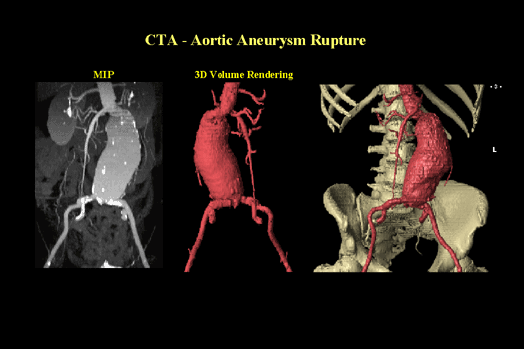
CTA - Aortic Aneurysm Rupture (Figure 9) |
|
A helical CT was performed on this patient for a pulsatile mass. The CT exam shows a large, leaking abdominal aortic aneurysm inferior to the renal arteries and extending to the bifurcation. The maximum intensity projection provides additional information by displaying calcified plaque within the aneurysm and the iliac arteries. Note the exceptional view of the Superior Mesenteric Artery and its branches located just superior to the aneurysm. The 3D views help give relational information about the aneurysm to bony landmarks. Exams like this are easy and quick to perform. They require little or no editing and can be completed in just a few minutes.
|
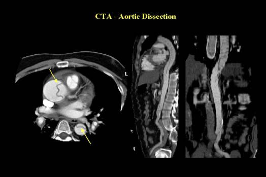
CTA - Aortic Dissection (Figure 10) |
|
This patient was involved in an automobile accident and transported to the ER. Helical CT shows a large dissection of the aorta extending from the arch to the descending aorta. The original helical image on the left shows the dissection clearly. The upper arrow is pointing to the dissection in the arch and the lower arrow is pointing to the dissection in the descending aorta. As a side note, the tricuspid valve is clearly shown as are some of the coronary arteries. The multiplanar reformats in the sagittal and coronal views help define the extent of the dissection. In cases such as this breath-hold is usually not possible but by virtue of the short scan times excellent image quality is possible.
|
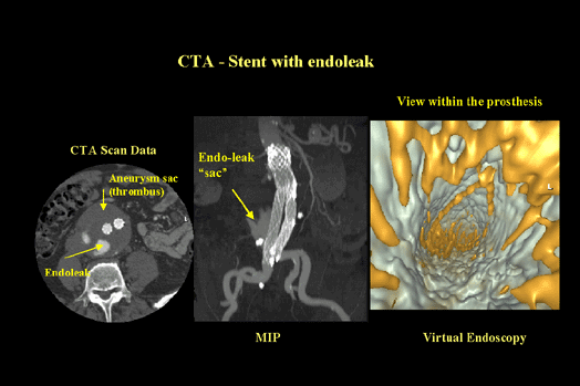
CTA - Stent with endoleak (Figure 11) |
|
This helical CT exam was performed on a patient with an abdominal aortic aneurysm that has undergone a stent placement. The examination shows a large thrombosed aneurysm with two stents. Posterior to the stents is a leak. The original image clearly shows the stents and the endoleak. The maximum intensity projection gives an overall perspective of the vasculature, stents and leak. The image on the far left is a view from within the stent known as virtual endoscopy. You can see the mesh of the stent and the outside wall of the vessel. This view is the same as you would use for the colon, airways or sinuses.
|


