|
The fast acquisition of thin overlapping slices with the simultaneous evaluation of vasculature, soft tissues and bony structures makes helical CT ideal in the evaluation of trauma patients. Clinical situations that in the past required the use of multiple time consuming modalities can now be evaluated quickly with a rapid helical CT examination. Current scanners allow for rapid evaluation from head to toe. (Figures 2 & 3)
|

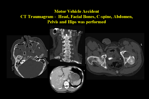
Motor Vehicle Accident (Figure 2) |
|
This examination is on a single patient and is referred to as a traumagram. A major advantage of helical CT is its ability to scan the whole body rapidly to assess trauma and get the patient to the next point of treatment without delay. Moving clockwise from the left: A head CT for facial bones demonstrates air and fluid, probably blood, within the maxillary sinuses. Note the depressed fracture on the right side as well as swelling of the nasal septum. Coronal reconstruction of the C-spine demonstrates all the cervical vertebrae in alignment. Special note: C-1 and C2, also referred to as the atlas and axis, are clearly demonstrated. Pelvic trauma showing dislocation of the left femoral head from the acetabulum. This CT of the abdomen demonstrates a ruptured spleen with contrast pooling postero-medially to the spleen.
|

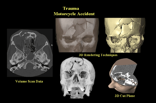
Trauma Motorcycle Accident (Figure 3) |
|
CT examination of the facial bones and cranium shows multiple depressed fractures. Moving clockwise from left: Axial image, using Reid's baseline, shows a depressed supraorbital ridge on the right as well as fluid within the sinuses. 3D reconstruction of the soft-tissue and bone. Note that the soft-tissue has a translucency applied to show the bony structures beneath. This is an excellent way to show surface landmarks in relation to the underlying structures. 3D reconstruction of the skull shows multiple depressed fractures. 3D reconstruction using a cut plane permits the diagnostician and surgeon to "melt away" overlaying structures to view internal detail. This image shows surface landmarks in relation to the fluid-filled sinuses. The final image is a MIP image of the bony detail. This image helps define the relationships of the fracture fragments. Note the clear depression in the frontal bones as well as the displaced fracture fragments in the right orbit.
|
CT is the method of choice when imaging for head and abdominal trauma (Figure 4). CT is best for assessing the extent of soft tissue injury and identifying entrance and exit wounds. For thoracic trauma, CT can detect and assess sites of injury to the lungs, including pulmonary embolus, contusion and laceration as well as injuries to the chest wall. Conventional x-ray is the primary exam of choice for patients with severe spinal trauma, but CT is the method of choice for studying the integrity of bony canal and identification and localization of bony fragments. (Figure 5)
|

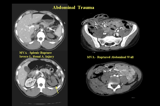
Abdominal Trauma (Figure 4) |
|
Abdominal trauma can easily be assessed using CT. The images on the left show a patient with a ruptured spleen as well as an injury to the left renal artery. In the image showing the splenic rupture, there is fluid collecting posteriorly to the spleen. The image displaying the left renal artery injury shows the kidney on the right receiving contrast while the left kidney is void of contrast enhancement. The images on the right show a severe trauma to the lower abdomen. The top image shows intestine spilling out through a laceration in the abdominal wall. The image below that shows pooling of blood into the soft-tissue on the right which is the result of a severed iliac artery.
|

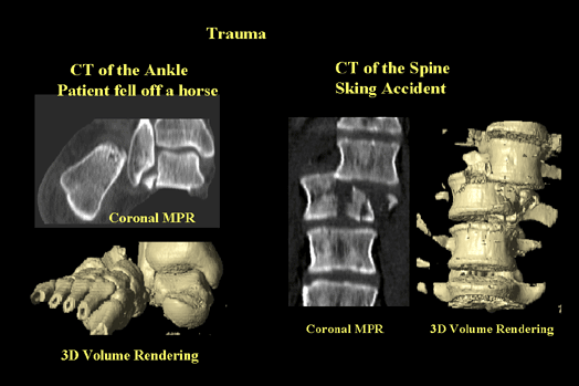
Trauma (Figure 5) |
|
A combination of reconstruction techniques including multiplanar reformation and 3D displays helps identify relational information. The two images on the left show a fracture and dislocated right foot. The coronal MPR gives a clear view of the tibia, fibula and talus with a fracture fragment in the space between the fibula and talus. A portion of the calcaneous is displaced laterally. A 3D image shows the extent of the bony injury and will help the surgeon define their surgical approach. The case on the right shows a severe spinal fracture with multiple vertebral body fractures and dislocation of the superior vertebrae. The coronal MPR gives an excellent view of the fractures as well as the dislocation. A 3D shaded surface display will help the surgeon define the surgical approach and stabilization methods.
|