風蕭蕭_Frank
以文會友


腦細胞能否再生不重要,有案例證明,整個左半腦切除,人的生活能力基本能恢複到正常。MRI掃描發現,在右腦的某些區域,發展為代替右腦的功能。
下麵是從我的文章摘錄複製的:
1.2.. A 32 years old man recovered as normal after lost left brain
http://www.kwcg.ca/bbs/home.php?mod=space&uid=61910&do=blog&id=6002
Dec. 14, 2015, YouTube published an episode of video The secret of a man with a half brain with title in Mandarin. https://www.youtube.com/watch?v=BqSmSBPmsik
The video introduced a British man Mr. Charie, in 2012, 32-year-old Charie travels in Thailand, fell down from 7 meters high with head to the ground, his all signs of life disappeared, 1/4 skull crushed, the left brain removed.
After two days of surgery, he miraculously recovered, after a period of language barriers and obstacles to action, he recovered as normal.
The doctor implanted a Titanium plate as skull into his brain and fixed with screws on the remained skull. His appearance and body flexibility were recovered back to normal.


Above were the photos of Mr. Charie from video screenshots
In 2015, 35 years old Mr. Charie has a job, lives in a normal life with his family in the UK.
1.4.. A man lives with half of brain
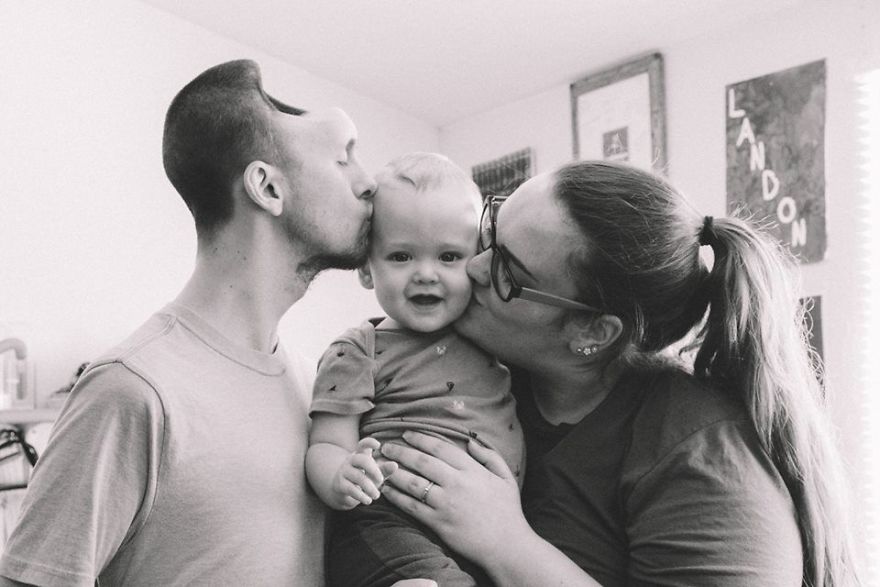
October 13, 2017, article Woman Shares What Energy Drinks Did To Her Husband While She Was 9 Months Pregnant reports a man's brain was damaged by Energy Drinks, after surgery and two weeks of living in a hospital, he survived back home.
Follow was photo of the happy family.
1.1.. The lost functions with cut of left brain developed on remained right brain
Aug. 14, 2917, YouTube published an episode of video Discovery Brain Nerves with title in Mandarin.
https://www.youtube.com/watch?v=9jDd0lF5bak&t=1008s
The video introduced a computer enhanced brain image of a 35 year old man Mr. Boo, when was as a boy, he underwent the surgery of cut off left brain.
Now, Mr. Boo lives a normal life.
Follow was the video screenshots of a PET scan of the right brain of Mr. Boo.

The colors reveals that his speech ability normally handled by the brains left side has migrated to the right side where Boo’s speech ability handled by other neural networks.
Sorry, Adults, No New Neurons For Your Aging Brains
Study questions whether adults can really make new neurons
Birth of New Neurons in the Human Hippocampus Ends in Childhood
Human Brains Stop Creating New Neurons in Adulthood
大腦不斷出現新生細胞 凸顯生命奧妙無限
2016-03-18 9:09 AM
http://www.epochtimes.com/gb/16/3/15/n4663356.htm?
科學家首次觀察到大腦中新生腦細胞的具體活動情況,這些細胞和學習記憶等認知能力有關。該研究進一步證實,成年大腦會產生新的腦細胞,大腦在出生後的細胞數量不是固定不變的。
最新一期《神經元》(Neuron) 雜誌登載的一項研究報告描述,科學家首次在活的小鼠大腦中觀察到新生腦細胞發射信號的情形。
而且這些新生腦細胞(也稱為“神經元”)和動物的學習與記憶有關係。如果這些新生神經元的功能被屏蔽,那麽動物出現學習和記憶障礙而不能很好的適應周圍環境。
據《新科學家》近日報導,長期以來,我們一直認為大腦的細胞數量在出生後就已經固定,腦細胞隻能隨年齡增長而越來越少。但是現在我們改變了看法,因為最新研究在大腦的具體區域看到了新生腦細胞的活動情況。
哥倫比亞大學等研究機構的科學家首次使用雙光子顯微鏡,在活的小鼠大腦齒狀回(dentate gyrus)中找到新生神經元在活動時發出信號的情況。科學家認為齒狀回區域和學習記憶有關。
新生腦細胞和認知功能有關
在成年鼠的這個大腦區域,新生和已經成熟的神經元相互混雜。該成像方法顯示出新生的神經元發出紅色螢光,而那些出生前即已經成熟的神經元發出綠色螢光。
報導說,科學家讓小鼠在跑步機上運動,同時給它不同的氣味(如香蕉的氣味)、聲音、光線等刺激。有時給小鼠的是它喜歡的東西,有時是能讓它厭煩或害怕的刺激甚至電給予擊,之後小鼠會形成各種記憶。
研究人員發現,小鼠在對待那些刺激時,其大腦中的新生神經元比成熟神經元的反應更活躍、更容易興奮、發射的信號更散亂。但是,這種活躍興奮的持續時間不長。而六周後,新生神經元的功能變得更加成熟。
研究人員尤其注意到,如果抑製這些新生腦細胞的功能,小鼠則不能很好地記憶在跑步機上的經驗,而且對周圍的新情況適應力變差,例如有感到更害怕等表現。
大腦不斷產生新的腦細胞
報導說,該研究至少證實成年大腦中的新生神經元對於學習是非常關鍵的因素,這一研究可能有助於尋找抑鬱、焦慮等疾病的治療方法。
德克薩斯州立大學的神經學家麥克?德魯(Michael Drew)對此研究評述到,該實驗非常好,給人留下深刻的印象,因為能觀察到活的神經細胞的功能是很不容易的。
早在2012年11月,紐約石溪大學(Stony Brook University)發現成年動物的大腦有新生神經細胞,即使已經衰老的大腦也會產生新的腦細胞。
石溪大學的科學家是在大腦的海馬體中發現這些細胞。因為這個區域和學習記憶有關,所以認為大腦新生細胞和學習等認知能力有關。責任編輯:林妍
Newborn neurons -- even in the adult aging brain -- are critical for memory
https://www.sciencedaily.com/releases/2012/11/121111152944.htm
 Dr. Shaoyu Ge and Yan Gu of Stony Brook University display the image of newborn neurons illustrated by the new optogenetic labeling and controlling technique.Credit: Image courtesy of Stony Brook University
Newly generated, or newborn neurons in the adult hippocampus are critical for memory retrieval, according to a study led by Stony Brook University researchers to be published in the November 11 advanced online edition of Nature Neuroscience. The functional role of newborn neurons in the brain is controversial, but in "Optical controlling reveals time-dependent roles for adult-born dentate granule cells," the researchers detail that by 'silencing' newborn neurons, memory retrieval was impaired. The findings support the idea that the generation of new neurons in the brain may be crucial to normal learning and memory processes.
Previous research by the study's lead investigator Shaoyu Ge, PhD, Assistant Professor in the Department of Neurobiology & Behavior at Stony Brook University, and others have demonstrated that newborn neurons form connections with existing neurons in the adult brain. To help determine the role of newborn neurons, Dr. Ge and colleagues devised a new optogenetic technique to control newborn neurons and test their function in the hippocampus, one of the regions of the brain that generates new neurons, even in the adult aging brain.
"Significant controversy has surrounded the functional role of newborn neurons in the adult brain," said Dr. Ge. "We believe that our study results provide strong support to the idea that new neurons are important for contextual fear memory and spatial navigation memory, two essential aspects of memory and learning that are modified by experience.
"Our findings could also shed light on the diagnosis and treatment of conditions common to the adult aging brain, such as dementia and Alzheimer's disease," he said.
Dr. Ge explained that their findings may help specifically to advance research on cell-replacement therapies being investigated for neurological diseases of the brain affecting memory.
In the study, Dr. Ge and colleagues used a retroviral tool to deliver optogenes: genes that are engineered to express proteins that form channels responsive to light stimulation. Using their created opto-retrovirus, the research team labeled a cohort of newborn neurons that they could control with light illumination. By doing this, the team conducted an in-depth exploration of the circuit and behavioral functions of newborn neurons in the adult mouse brain.
The researchers first determined when the neurons were "ready" in the hippocampus, which is an important addition to their findings published earlier this year in Nature. Then they 'silenced' the activity of neurons of different ages. They found that by silencing four-week-old neurons, an age known to be more responsive to change than existing neurons, but not older or younger neurons, memory retrieval was impaired in tasks known to depend on the functions of the hippocampus.
Regarding the newborn neuron controlling method, Dr. Ge and colleagues said that the novel tool and approach used in the study can be applied to other circuit and behavioral studies to access the functional contributions of newborn neurons in the adult brain.
Co-authors of the study include: Yan Gu, Jia Wang, and Stephen Janoschka of the Department of Neurobiology and Behavior at Stony Brook University School of Medicine; and Maithe Arruda-Carvalho, Sheena Josselyn, and Paul Frankland of the University of Toronto in Canada.
Story Source:
Materials provided by Stony Brook University. Note: Content may be edited for style and length.
Journal Reference:
Yan Gu, Maithe Arruda-Carvalho, Jia Wang, Stephen R Janoschka, Sheena A Josselyn, Paul W Frankland, Shaoyu Ge. Optical controlling reveals time-dependent roles for adult-born dentate granule cells. Nature Neuroscience, 2012; DOI: 10.1038/nn.3260
Cite This Page:
Stony Brook University. "Newborn neurons -- even in the adult aging brain -- are critical for memory." ScienceDaily. ScienceDaily, 11 November 2012.
- November 11, 2012
- Stony Brook University
- Newborn neurons in the adult hippocampus are critical for memory retrieval, according to a new study.
研究再次發現 成熟大腦細胞可再生
http://www.epochtimes.com/gb/17/8/21/n9552207.htm
科學家再次在成熟大腦中發現神經細胞再生現象,這可能有助於治療焦慮、抑鬱和創傷後應激障礙(PTSD)等心理疾病。
澳洲昆士蘭大學的科學家近日發表報告說,在成熟小鼠大腦的杏仁核(amygdala)裏發現新生腦細胞。杏仁核與處理情緒和記憶有重要關係,該區域損傷或有病變,可能會發生焦慮症,如創傷後應激障礙。
昆士蘭腦研究所主任薩赫(Pankaj Sah)表示,該新發現對於進一步了解大腦的適應和再生能力有重要意義。
薩赫教授說:“現在,我們在成年小鼠大腦的杏仁核中發現幹細胞,這表明在海馬和杏仁核兩側有新生神經細胞的現象。該發現加深了我們對腦可塑性的理解,為研究杏仁核的新生神經細胞的功能和分布提供思路。”

研究者之一加弗利(Dhanisha Jhaveri)博士說,杏仁核在恐懼學習中發揮了關鍵作用。
加弗利博士解釋:“恐懼學習會導致經典的逃離或打鬥反應,如心率增快、口幹、手掌出汗。但是,在恐懼症或創傷後應激障礙的病例中,杏仁核也會產生恐懼和絕望的感覺。”
因此,科學家根據該研究,希望能找到刺激杏仁核產生新細胞的方法,以便嚐試治療焦慮、PTSD和抑鬱症的新療法。

“雖然以前知道成熟大腦中會產生新神經元。但這是在杏仁核中首次發現新生細胞,令人非常高興。”薩赫教授說,“該發現對人們理解杏仁核在調節恐懼和恐懼記憶中的作用有著重大意義。”
昆士蘭大學的科學家巴特利特(Perry Bartlett)教授曾在海馬中發現神經細胞再生的現象。海馬是大腦中與空間學習和記憶有關的重要腦區。
近一百年來,科學界認為成熟大腦無法再生細胞,腦細胞隻能不斷地發生死亡。薩赫教授表示,巴特利特教授的發現推翻了人們認為大腦細胞不可再生的看法。
巴特利特教授也參與了此次的最新研究。
日實現活體動物腦內神經細胞再生
新華社東京7月16日電日本研究人員15日在英國《自然雜誌神經學專刊》網絡版上報告說,他們首次在活體實驗鼠腦內實現神經細胞再生。這一成果有望促進神經再生醫療研究。
活體動物腦內神經細胞再生
此前科學界一直認為,可生成腦內神經細胞的幹細胞,其功能在胎兒時期就基本停止,即使出生後由於事故和疾病導致腦損傷,其腦神經幹細胞也無法發揮再生作用。
日本東京大學的研究人員發現,在胎兒的腦神經幹細胞中,高遷移率族蛋白A一直在發揮作用,但該蛋白在嬰兒出生後很快就不發揮作用了。研究小組選取了能使這種蛋白持續發揮作用的基因,並將其植入出生僅數天的實驗鼠的大腦神經幹細胞,結果實驗鼠恢複了腦神經細胞再生能力。
領導這項研究的後藤由季子教授表示,下一步他們會利用成年實驗鼠測試這種方法能否實現神經細胞再生
《自然》:死一個少一個!中美科學家聯合發現,成年人腦細胞無法再生,珍惜吧 | 科學大發現
2018-03-09 08:20 奇點網
https://3g.163.com/tech/article/DCEK1NEI051182D7.html
在過去的幾十年裏,不斷有新的證據表明,人類大腦神經元是可以自我更新的,這無疑給治療和預防神經係統疾病帶來了希望。不過這個希望在今天被暫時打破了。本周《自然》上的一項新研究表明,人類大腦中新生神經元的數量會隨發育急劇下降,而成年人則不會產生新的神經元[1]。
真的是個悲傷的消息,這意味著我們的腦細胞用一個少一個,一傻不複還了。
 上:豐富的新生神經元(綠)下:成年後則不產生新生神經元
對於人類大腦中新生神經元的爭論是自古就有的。神經科學剛剛發展起來的時候,學界的普遍共識就是,大腦神經元像心肌等細胞一樣,出生的時候是多少就隻有多少,不會進行自我修複和更新。
不過到了20世紀60年代,一項研究打破了這個定論。當時麻省理工的Joseph Altman發現,在成年哺乳動物的大腦內存在新生神經元。又過了一段時間,80年代,洛克菲勒大學Fernando Nottebohm實驗室也在禽鳥身上發現了類似的現象。1998年,科學家們得到了第一份證據,在成年死亡癌症患者腦中發現了由5-溴脫氧尿苷(BrdU)標記的新生神經元。2013年的一項研究更是令學界大振——通過分析55名死者腦組織的單個神經元,卡羅林斯卡研究所的研究者們得出了一個結論,人類大腦海馬齒狀回中,每天都能產生700個新的神經元。[2,3]
海馬是人類大腦中的一個關鍵區域,它已經被證實,和學習、記憶、壓力、鍛煉等多個生理過程有關,它的變化涉及到各類神經係統疾病[4-8]。如果大腦可以通過產生新的神經元進行自我更新,那麽提高先天再生能力來恢複受損大腦的活動就變成了一條可行的路。
本項研究的通訊作者之一,就職於加州大學洛杉磯分校的Arturo Alvarez-Buylla也是致力於這個領域的研究者之一。當年他還是Fernando Nottebohm實驗室的一名普通學生,而今天已經成為了腦發育領域的頂尖專家。過去的三十年裏,他對大腦新生神經元科研做出了極大的貢獻。他也堅信,在成年人類大腦中也存在新生神經元。

Arturo Alvarez-Buylla教授
然而事實並非如此。
鑒於人類大腦標本實在難以獲得,Alvarez-Buylla教授組建了一個國際團隊,最終獲得了59份人類腦組織標本,分別來自屍檢樣本和癲癇手術切除的腦組織樣本,其中成年人的年齡層從18-77歲。這項工作我國複旦大學腦科學研究所的楊振剛教授也有參與,並名列通訊作者之一。
研究者對這些腦組織樣本進行了抗體熒光標記,並通過影像學技術確認細胞形態來驗證。結果顯示,腦部的新生神經元主要出現在胎兒發育期,並在14周的時候數量達到頂峰,22周以後則開始減少;同時,新生神經元表現出細長而簡單的細胞形態,並隨著發育逐漸生長為成熟的神經元形態。而到了7歲左右,就基本不會再產生新生神經元了。
研究者對各年齡段的海馬顆粒細胞層(GCL)新生神經元進行了計數。在剛出生的時候,每平方毫米的新生神經元能夠達到1618±780個;在1歲的時候,下降到292.9±142.8;7歲時,僅有12.4±5.3;13歲就隻有2.4±0.74這麽一點點了。
而在成年人的腦標本中,研究者沒有發現任何分裂細胞/新生神經元的跡象。

隨年齡增長,新生神經元逐漸減少(綠)
研究者也嚐試在恒河獼猴腦中尋找分裂細胞和新生神經元的蛛絲馬跡。
關於恒河獼猴腦中新生神經元是否存在,既往的兩項研究采取了不同的標記方式,也產生了截然不同的結果[9,10]。研究者用研究人腦標本一樣的思路對不同年齡段的猴腦進行了檢測,發現在出生後到一歲半期間,新生神經元的數量降為1/8;在一歲半到七歲之間,更是直降為1/35。新生神經元的數量變化大體上與人腦一致。

使用兩種標記方式標記的猴腦中新生神經元
最後,研究者檢測了人腦和猴腦在一歲半時的表達譜,結果顯示與新生神經元相關的DCX、TUJ1、Ki-67等蛋白的表達顯著降低。

左中右分別為分裂細胞的標記、新生神經元的標記、成熟神經元的標記
從整體結果來看,毫無疑問,我們能夠得出結論,海馬的新生神經元數量在出生後顯著下降,並且成年之後基本不會產生新神經元。但是這項研究成果與以往的一些數據既然相反,在同領域的研究者之間引起了爭議。
德國德累斯頓科技大學的神經科學家Gerd Kemperman表示,隻是科學家沒有觀察到新的神經元,這並不意味著新神經元不存在。研究中使用的腦組織畢竟隻是樣本,對新生神經元標記的可靠性很大程度上取決於組織的質量,很容易受外部環境的影響。
阿姆斯特丹大學的神經科學家Paul Lucassen也表示,用於保存和穩定組織樣本的化學物質也可能進一步防止標記和靶細胞的結合。
另外一些神經科學家還提出,腦組織的主人身體和神經狀態也是很重要的。運動、壓力、疾病等因素都會影響新生神經元的數量[11]。

對於這些疑問,Alvarez-Buylla教授表示認同,並提出了自己的看法。既往研究的標記方法有可能混淆了新生神經元和膠質細胞,因為它們會產生同樣的標誌物;另一方麵,腦組織受到死亡情況和疾病情況的影響,但是在具有多種死亡原因和疾病狀態的患者組織都得到了類似的結果,這也能夠一定程度上說明問題。
研究者承認這項研究的局限性——再怎麽仔細地檢測,也不能夠完全說明海馬中不存在新生神經元。但是第一作者之一的Shawn F. Sorrells也提出了一個觀點:“我們退一步想,如果新生神經元已經罕見到我們無法檢測,那麽它真的在海馬的記憶和學習功能中發揮重要作用嗎?”
樂觀一點想,發現成人海馬不存在新生神經元,也未必是件壞事。目前我們已知齧齒類動物和禽類成年後仍會產生新的神經元,而海豚和鯨類這些更加聰明、行為更加複雜的水生動物則也不會產生新生神經元[14]。知曉了這些差異,對於我們進一步研究這些差異為何產生是很有幫助的。
畢竟科學就是個探索未知的過程嘛。
參考資料:
[1] https://www.nature.com/articles/nature25975#f14
[2]http://www.cell.com/cell/fulltext/S0092-8674(16)31404-0
[3] Spalding, K. L. et al. Dynamics of hippocampal neurogenesis in adult humans.Cell 153, 1219–1227 (2013).
[4] Kempermann, G., Kuhn, H. G. & Gage, F. H. More hippocampal neurons in adult mice living in an enriched environment. Nature 386, 493–495 (1997).
[5] van Praag, H., Kempermann, G. & Gage, F. H. Running increases ce llproliferation and neurogenesis in the adult mouse dentate gyrus. Nat. Neurosci.2, 266–270 (1999).
[6]Lugert, S. et al. Quiescent and active hippocampal neural stem cells with distinct morphologies respond selectively to physiological and pathological stimuli and aging. Cell Stem Cell 6, 445–456 (2010).
[7] Malberg, J. E. & Duman, R. S. Cell proliferation in adult hippocampus is decreased by inescapable stress: reversal by uoxetine treatment. Neuropsychopharmacology 28, 1562–1571 (2003).
[8] Hill, A. S., Sahay, A. & Hen, R. Increasing adult hippocampal neurogenesis is sufcient to reduce anxiety and depression-like behaviors. Neuropsychopharmacology 40, 2368–2378 (2015).
[9] Kornack, D. R. & Rakic, P. Continuation of neurogenesis in the hippocampus of the adult macaque monkey. Proc. Natl Acad. Sci. USA 96, 5768–5773 (1999).
[10] Gould, E. et al. Hippocampal neurogenesis in adult Old World primates.Proc. Natl Acad. Sci. USA 96, 5263–5267 (1999).
[11] http://dx.doi.org/10.1007/s00441-017-2735-4
[12]https://www.nature.com/articles/d41586-018-02812-6
[13]https://www.sciencedaily.com/releases/2018/03/180307141356.htm
[14] Knoth, R. et al. Murine features of neurogenesis in the human hippocampus across the lifespan from 0 to 100 years. PLoS ONE 5, e8809 (2010).
成年人神經或無法再生
作者:宗華 來源:科學網 2018/3/9 18:50:04

新神經元在新生兒的大腦內是可見的(上圖),但並未出現在35歲成年人的大腦組織中(下圖)。
圖片來源:KEN PROBST
過去20年間,成年人每天能產生上百個新神經元的證據,點燃了增強治療性細胞誕生能力的希望。研究人員推斷,促進神經再生或能預防或者治療抑鬱症、阿爾茨海默氏症和其他大腦疾病。不過,一項日前發表於《科學》雜誌的爭議性研究表明,神經元的產生在經曆早期發育後大幅減少並且到成年時慢慢停止。
加拿大多倫多病童醫院神經科學家Paul Frankland表示,在成年人和猴子大腦中“竭盡全力搜尋”新神經元的結果“將會令很多人失望”。美國哥倫比亞大學神經科學家René Hen則表示,“最新研究引發了這樣的擔憂,即神經再生水平過低,以至於無法在人類體內產生重要功能。”
1998年,關於成年人神經再生的首個證據來自死亡的癌症患者的大腦。他們在世時,曾接受過一種名為溴脫氧尿苷的化學物質的注射。該物質能標記新分化的細胞,並且出現在這些患者大腦內的一些海馬體(和記憶、學習相關的海馬狀結構)中。2013年,來自瑞典卡羅林斯卡研究所的Jonas Frisén團隊對55名已去世患者大腦組織中的單個神經元進行了碳年代測定。該團隊根據細胞年齡推測,人類每天在編碼記憶的一個海馬體區域——齒狀回區更新700個神經元。
不過,自上世紀80年代起便一直研究大腦產生新細胞能力的加州大學舊金山分校研究人員Arturo Alvarez-Buylla對此持懷疑態度。他帶領團隊花費5年時間,收集了來自不同年齡段(從出生前到77歲)的59人的大腦組織。這些人當時已經死亡,或者其大腦組織在癲癇手術期間被移除。研究人員利用熒光抗體標記了對處於不同成熟度的細胞具有特異性的蛋白。他們還利用電子顯微鏡,尋找了特有的細長且形狀簡單的年輕神經元。
該團隊發現,人們在生命早期擁有大量的神經幹細胞和祖細胞——出生時,每平方毫米大腦組織平均擁有1618個年輕神經元。不過,這些細胞並未繼續形成神經幹細胞的增殖層,同時新神經元的產生在1~7歲減少了23倍。到成年時,新神經元的供應逐漸完全消失。(宗華)
m
